|
Elaine Sbroggio de Oliveira
Rodini, Dr and Aguinaldo Robinson de Souza, Dr
I believe that society will collapse unless
the average competence of its members for maintaining life support systems
is greatly increased, and unless the capabilities of its more talended
members are fully developed in terms that enrich and support the general
good" Potter, V.R.: Bioetics for whom?
Ann. N.Y. Acad. Sci. 196/4:200-205, 1972
I - General Aspects of Down Syndrome
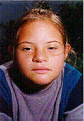 Fig. 1. |
Down Syndrome (DS) is the most
well known genetic syndrome . Fifteen percent of patients institutionalized
for mental retardation have DS. The features of DS was first reported by
Langdon Down in 1866. This condition is also called mongolism , because
the facial appearence of the patients (fig. 1).
The correct diagnosis is made by the karyotype,
that is the representation of the chromosomes of a cell. In the figure
2 is showed a karyotype of a DS patient. The karyotype is usually performed
through leukocytes from a peripheral blood sample. It is possible to realize
the karyotype before birth, after twelfth gestational week, using fetal
tissue. |
 Fig. 2. karyotype of a DS patient |
Recently has been used a ultrasonographic marker that may suggest DS diagnosis in twelfth gestational week. This marker is known as nuckal translucence, and it is a measure obtained from fetal nuckal region. The values above 3 mm suggest presence of congenital defects (presenting at birth), among then, the DS. In this cases has been indicated fetal chromosomal study (2). The incidence of DS is nearly 1/800 live births.
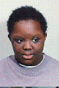 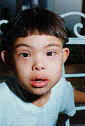 Fig. 3.Patients with DS,a black girl and a japanese boy |
There is a relevant relation between
the conception of DS children and maternal age. After 35 years of age,
mothers have increased risk to concept children with DS. When mother is
20 years old, the risk is 1/1600, while at 35 this risk is 1/370. DS occurs
in all races and in both sexes. On figure 3 are showed two patients with
DS,a black girl and a japanese boy.
Clinical features of DS are congenital and include meanly: mental retardation, muscle hypotonia (weaknees), ahort stature, cardiac anomalies, flat profile (fig.4), short and low set ears (fig.5), upslanting palpebral fissures (fig.6), protruding, broad and furrowing of tonge (fig.7), curved fifth finger (fig.8), gap between hallux, second toe single palmar crease (fig.9), and single palmar crease (fig.10). |
Characteristics of Down Syndrome
 Fig. 4 flat profile |
 Fig. 5. short and low set ears |
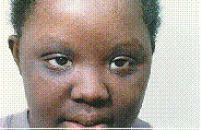 Fig. 6. upslanting palpebral fissures |
|
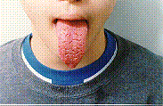 Fig. 7. broad and furrowing of tonge |
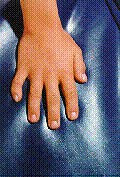 Fig. 8. curved fifth finger |
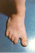 Fig. 9. gap between hallux and second toe |
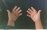 Fig. 10. single palmar crease |
The child with DS must be conducted so earlier
as possible to the particular services that will guide parents about prognosis
and specific treatment. The life quality of the patients depends on family
caring. Early stimulation improves the neuro-motor development, muscle
hypotonia and speech (7).
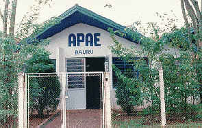
Specialized institutions for DS as APAE offer specific programs for early stimulation. Others examples include the Fundação Sindrome de Down (http://www.aleph.com.br/cdi/) with home office in Brazil, and the Down Syndrome WWW Page (http://www.nas.com/downsyn/) with home office in USA.
The average life expectancy of the DS patients is 35 years, and depending of the presence and gravity of cardiac disease. Fertility in man with DS is severely reduced due mental retardation. About 50% of cases, the offspring has DS.
DS patients presenting declining imunologycal function, loading them to infections and increasing risk to develop cancer, particulary leukemia (6). Respiratory diseases are common. Aproximately 65 to 80% of all fetuses with DS are lost spontaneously (1).
II - DS Etiology
The excess of genetic material from chromosome 21: free trysomy in all cells of the body, chromosomal translocation, and free trisomy in part of the cells of the body ( Mosaicism ).
II.1 - Free Trisomy in all cells of the body
Fig. 11. representation of three chromosomes 21. |
Nearly 92% of DS patients have a extra chromosome 21 in all of their cells, resulting in a karyotype with 47 chromosomes, due to trisomy 21. |
The genetic mechanism that results in free trisomy is the non-disjunction of the pair 21 during gametogenesis (meiosis) from one of the parents resulting in a sperm or ovule with 24 chromosomes, due to disomy (two chromosomes) 21. After fertilization is originated a DS embryo. Non disjunction is more frequent in woman, meanly after 35 years of age. If a couple had a DS child due to free trisomy, the reccurrence risk for another child is estimated on 1%. DS child siblings do not presenting incresead risks to have affected childreen.
II.2 - Chromosomal translocation
Fig. 12. Pair 14, with a translocation |
In 3 to 4% of DS cases, chromosomes 21 extra
is linked to another one, usually chromosome 14. This rearranjement is
named translocation. In the figure 12 is showed the pair 14, with a choromosome
21 linked.
The karyptype, in this case, presents 46 chromosomes and the translocation is represented as t(14;21) or t(14q21q). The letter q refers to long arm of the involved chromosomes. These translocations may be balanced, if there is not an excess of chromosomal material, or unbalanced, if there is an excess. In figure 13 are showed a pair of chromosome 21 and a extra chromosome 21 linked to the long arm of chromosome 14. |
 Fig. 13. Pair of chromosome 21 and a extra chromosome 21 linked to the long arm of chromosome 14. |
The DS child parents may have a balanced translocation, presenting 45 individualized chromosomes, but with a genetic material corresponding to 46 chromosomes, because one of the two chromosomes from pair 21 is linked with another chromosome. When is the mother that presents this translocation there is a risk of 12% to have another child with DS, and, when is the father, the risk is 3%. The reason for that remains unclear. Is a child has DS due to translocation, it is important to perform the karyotype of her/his parents. |
In about ¾ of the DS patients the translocation
is not present in the parents but results from an error during parents
gametogenesis, origining a translocated ovule or sperm. In these cases
the reccurrence risk to another child is 2 to 3%. DS patients due to translocation
are indistinguishable from those with free trisomy. There is no relation
betwenn chromosomal translocation and maternal age.
II.3 - Mosaicism of the Chromosome 21
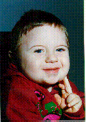 Fig. 14. child with mosaicism of chromosome 21 |
Mosaicism of the chromosome 21 is present in 2 to 4% of the DS patients. They presents two kinds of cells, one with normal number of chromosome (46), and anothert with abnormal (47). The mean cause of mosaicism is the non-disjunction of chromosome 21 during mitosis ( somatic cells division ) in embryo. A cell with non-disjunction origins trisomyc cells. The result is a combination of normal and abnormal cells. As low the number of trissomic cells, lower is the phenotypic involvement. Usualy mosaic patients are less affected. In figure 14 is showed a child with mosaicism of chromosome 21. This form of DS is not related with maternal age (3). |
III - Brain features of DS patients
The more frequent feature in DS patients is mental retardation. Brain development is delayed, so that infants are microcephalic at birth. A decrease in total brain weight has been observed, besides simplification of gyriform patterns. Neuropathologic axamination has demonstrated that the cerebellum appear to be smaller than normal. Specific deficts have been documented in certain areas, includind audictory, visual, mind and language abilities. Atrophic changes characteristic of Alzheimer disease has been documented in older patients (5). IV - References
Authors:
 Elaine Sbroggio de Oliveira Rodini, PhD. Biogeneticist, master and doctor in Sciences by Universidade Estadual Paulista - UNESP. Department of Biology at UNESP - Bauru/SP, Brazil. E-mail: elaine@bauru.unesp.br Aguinaldo Robinson de Souza,
PhD. Chemist, master and doctor by Universidade de São Paulo, and
post doctoral fellowship by the University of California at San Diego.
Chemistry Department - UNESP - Bauru/SP, Brazil. Acknowledgements: We must thank the staff, and parents, of APAE - Bauru/SP, the permision to the child's pictures publication.
|