
2 of 3
The Components of Basal Ganglia
 |
 |
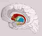
The principal anatomical units
of basal ganglia include:
![]() Caudate nucleus,
Caudate nucleus,
![]() Putamen,
Putamen,
![]() Globus pallidus
Globus pallidus
![]() Claustrum.
Claustrum.
The substantia nigra, and the subthalamic nucleus are generally included on a functional basis, while the amygdaloid body, located in temporal lobe, is functionallly excluded.
The major divisions of the basal
ganglia are:
![]() corpus
striatum is constituted by neostriatum (caudate
+ putamen) and paleostriatum (globus pallidus)- concerned with primarily
with somatic motor functions
corpus
striatum is constituted by neostriatum (caudate
+ putamen) and paleostriatum (globus pallidus)- concerned with primarily
with somatic motor functions
![]() amygdaloid
nuclear complex, functionaly related to the
hipothalamus and regarded as an integral part of the limbic system.
amygdaloid
nuclear complex, functionaly related to the
hipothalamus and regarded as an integral part of the limbic system.
|
|
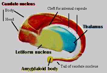
Constituted by: Situated between the insula, the caudate nucleus and the thalamus Its neurotransmitters include: Tracts conveying impulses from the globus pallidus and striatum to the substantia nigra contain GABA, while tracts carrying impulses from the substantia nigra to the caudate and putamen store dopamine in their terminals. This represents the most important source of dopamine in the striatum. Failure of dopamine syntesis in the substantia nigra provokes a progressive depletion of this chemical substance in the caudate and putamen, with a consequent development of Parkinson's disease. |
|
Lentiform nucleus |
Constituted by: The descriptive term "lenticular" or "lentiform" nucleus is applied to the putamen and globus pallidus togeather because of their combined lens-shape in brain sections. The globus pallidus can be divided into two parts:
the globus pallidus medial (GPm)and the globus pallidus lateral (GPl).
Both receive input from the caudate and putamen, and both are in communication
with the subthalamic nucleus. It is the GPm, however, that sends the major
inhibitory output from the basal ganglia back to thalamus. The GPl also
sends a few projections to the midbrain presumably to assist in postural
control. |
|
|
Although a component of the brainstem, the
substantia nigra
is considered along with the basal ganglia because it has reciprocal connections
with and is functionally related to the basal ganglia.
It can be divided into two parts: the substantia
nigra pars compacta (SNpc) and the substantia nigra pars reticulata (SNpr).
The SNpc receives input from the caudate and putamen, and sends information
right back. The SNpr also receives input from the caudate and putamen,
but sends it outside the basal ganglia to control head and eye movements.
The SNpc is the more famous of the two, as it produces dopamine, which
is critical for normal movement. The SNpc degenerates in Parkinson's disease,
but the condition can be treated by giving oral dopamine precursors. |
| Claustrum | Derived from a Latin word meaning a barrier.
is a thin sheet of gray matter.
It is located medial to the insular cortex and overlying the lateral surface of the putamen. It appears to have widespread reciprocal connections with sensory, particularly visual and somatosensory areas, of the cerebral cortex via association fasciculi in the extreme capsule. It also receives input from the hypothalamus (lateral), the thalamus (centromedian nucleus), and the locus ceruleus. Fibers from numerous cortical areas each terminate in distinct zones within the claustrum. Thus, it contains discrete somesthesic, visual and auditory zones. The claustrum has no subcortical projections. |
Although there are many different
neurotransmitters used within the basal ganglia (principally ACh, GABA,
and dopamine), the overall effect on thalamus is inhibitory. The function
of the basal ganglia is often described in terms of a "brake hypothesis".
To sit still, you must put the brakes on all movements except those reflexes
that maintain an upright posture. To move, you must apply a brake to some
postural reflexes, and release the brake on voluntary movement. In such
a complicated system, it is apparent that small disturbances can throw
the whole system out of whack, often in unpredictable ways. The deficits
tend to fall into one of two categories: the presence of extraneous unwanted
movements or an absence or difficulty with intended movements.
Author: Dr. Silvia Helena Cardoso, PhD.
Center for Biomedical Informatics
State University of Campinas, Brazil
Copyright 1997 State University of Campinas