
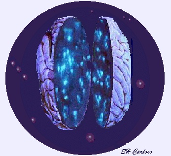
Caution: The image inside the brain are neurons, but they are not placed in this manner. Click here to see how the brain looks like in this view. |
Silvia Helena Cardoso, PhD Not only the stars in the Universe fascinate Man with its impressive numbers. In another universe, our own, biological one, a gigantic "galaxy" with billions of small neural cells forms our brain and the rest of the nervous system, and communicate among themselves by means of flashes of electrochemical pulses. They are resposible for everything: our feelings, thinking, emotions, pain, dreams, movements and sensations, and many other mental and physical functions. Without them, it would be impossible to achieve our rich internal world and to communicate with the surrounding environment, by means of sound, smell, taste, touch and light; including that of the stars in our Universe. |
.
Contents:

All stimuli of our environment causing sensations such as pain and hot, all feelings, thoughts, programming of motor and emotional responses, neural bases of learning and memory, actions of psychoactive drugs, causes of mental disorders, and any other action or sensation of the human being cannot be understanding without the knowledge of the fascinating process of communication between neurons.
Neurons are specialized
cells. They are made to receive certains specific connections, to perform
appropriate functions and pass their decision of a particular event to
other neurons which are also concerned with those events. These specializations
include a cell membrane,
which is specialized to convey nerve signals as electrochemical pulses;
the dendrite,
(from the Greek dendron, or tree) which gets and delivers
the signals, the axon
(from the Greek axoon, or axis), the conducting cable of electrical
signals, and points of synaptic contacts,
where information can be passed on from one cell to another (see fig.1).
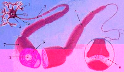
Fig.1. The structure of the neuron. A typical neuron has four morphologically defined regions: dendrites (1), cell body (2), axon (3), and presynaptic terminals (5). |
Neurons receive nerve signals from axons of other neurons. Most signals are delivered to dendrites (1). The signals generated by a neuron are carried away from its cell body (2), which contains the nucleus (2a), the storehouse of genetic information. Axons (3) are the main conducting unit of the neuron. The axon hillock (2b) is the site at which the cell's signs are initiated. Schwann cells (6), which are not a part of a nerve cell, but one of the types of glial cells, perform the important function of insulating axons by wrapping their membranous processes around the axon in a thight spiral, forming a myelin sheath (7), a fatty, white substance which helps axons transmit messages faster than unmyelinated ones.The myelin is broken at various points by the nodes of Ranvier (4), so that in cross-section it looks rather like a string of sausages. Branches of the axon of one neuron (the presynaptic neuron) transmit signals to another neuron (the postsynaptical cell) at a site called the synapse (5). The branches of a single axon may form synapses with as many as 1000 other neurons. |
See also: Parts of the Nerve Cell and Their Functions
What Make Neurons Different from Other Cells?
Just like other cells, neurons feed, breath, have the same genes, the same biochemical mechanisms and the same organelles. So, what makes the neuron different ? Neurons differ from other cells in one important respect: they process information. They must gather information about the internal state of the organism and his external environment, evaluate this information, and coordinate activities appropriate to the situation and to the person's current needs.
The information is processed through an event known as the nerve impulse.A nerve impulse is the transmission of a coded signal from a given stimulus along the membrane of the neuron, from the point that it was stimulated.Two types of phenomena are involved in processing the nerve impulse: electrical and chemical. Electrical events propagate a signal within a neuron, and chemical processes transmit the signal from one neuron to another or to a muscle cell. The chemical process on interaction between neurons occurs at the end of the axon, called synapse. Touching intimately against the dendrite of other cell (but without material continuity between both cells), the axon releases chemical substances called neurotransmitters, which attach themselves to chemical receptors in the membrane of the following neuron.
The
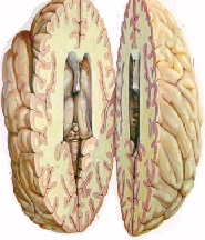
Brain is Grey and White. Why?
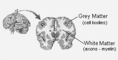
Maybe you have heard the term "grey matter" for the brain; there is also "white matter". In a section made through the brain, it is easy to see both grey and white areas. The cortex and other nerve centres are grey, the regions in between, white. The grey coloration is produced by the aggregation of thousands of cell bodies, while the white is the color of myelin. The white color reveals the presence of bundles of axons passing through the brain, rather than areas in which connections are being made. No neuron has direct conection with any other. At the far end of the axon are a number of terminal filaments, and these run up close to other neurons. They may be close to the dendrites of the other neuron (sometimes to special structures called dendritic spines, or close to the cell body itself. Where the first neuron comes close to the second neuron, a synapse is formed, a space acreoss which the first neuron communicate with the second. Next:Anatomical Diversity of Neurons
Resources
The
Brain and the Nervous System
Types
of Neurons
The
Biological Neuron
The
Neuron
Lights,
Camera, Action Potential!!
How
Animals Transmit Informations
Gallery
of Neurons
References:
Neuroscience:
Exploring the Brain. Bear, M.F.; Connors, Barry W. e Paradiso, Michael
(eds.)- Williams & Wilkins, 1996.
Human
Mind Expalined - A. A. Greenfield (ed) - Henry Holt And Company, 1996.
1
The Author
 Silvia
Helena Cardoso, PhD, Psychobiologist, Director and Editor-in-chief,
Brain & Mind.
Silvia
Helena Cardoso, PhD, Psychobiologist, Director and Editor-in-chief,
Brain & Mind.
Reviewed by the neuroanatomist Dr. Norberto Cysne Coimbra , MD, PhD. Laboratory of Neuroanatomy and Neuropsychobiology, School of Medicine of Ribeirão Preto, University of São Paulo (USP), Ribeirão Preto, Brazil