
Silvia
Helena Cardoso, PhD and Renato M.E. Sabbatini, PhD
Animation and Art: André Malavazzi
|
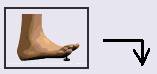
Fig.1.Simple
Reflex - Human feet in contact with a tack (external stimulus).
(Click on the button "Reinicia" ("Re-start") to see the animation). |
To
imagine how the nervous impulse happens, observe the figure on the left.The
perception of acute pain when a sharp object penetrates your foot is caused
by the generation of certain action potential in some nervous fiber in
the skin.
The skin perforation is transduced into signals that travel upwards via sensorial nerves (direction of information flow shown by arrows). This information arrives at the spinal chord and is distributed to inter neurons (neurons that provide intermediate connections with other neuron chains). Some of these neurons send axons to the sensorial region of the brain where the sensation of pain is registered. Other make synapses with motor neurons,which send signals downward to the muscles. The motor junctions command the muscle contraction and withdrawal of the foot. This is an example of a neuron chain called reflex arc. |
|
It
is believed that the membranes of these cells have sodium channels that
open when nerve terminals of the cell are stretched. The initial chain
of events is:
1.Sharp
object penetrates the skin;
Due to the concentration gradient and negative charge in extracellular fluid,ions enter the fiber by means of these channels. Sodium entrance(yellow balls) depolarizes the membrane, that is, the side of membrane bathed by extracellular fluid becomes less negative in relation to the cell interior. If this depolarization, called generator potential, reaches a critical point, the membrane then generates an action potential. The critical point of depolarization that must be overcome in order to fire an action potential is called threshold.The action potentials are caused by depolarization of the membrane above the threshold. In
the repolarization phase, the sodium channel is deactivated and the conductivity
of membrane for potassium increases. These positive ions
In the recovery phase, the active sodium/potassion pump (blue channel in the figure) restablishes the ionic levels which were altered during the action potential: for each sodim ion which is transported from the inside to the outside, a corresponding potassium is transported in the opposite direction. |

Silvia Helena Cardoso, PhD.Psychobiologist, master and doctor in Sciences, Founder and editor-in-chief,Brain and Mind Magazine, State University of Campinas. |

Renato M.E. Sabbatini, PhD.Doctor in Sciences (Neurophysiology) by the State University of São Paulo Associate Director of the Center for Biomedical Informatics of the State University of Campinas, Brazil, and president of the Editorial Board,Brain & Mind Magazine. |

André Malavazzi Designer, Instute of Arts and Center for Biomedical Informatics, State University of Campinas. |
Copyright (c) 2000 State University
of Campinas, Brazil
An initiative: Center for Biomedical
Informatics
Published: 15.Jan.2000