 Cogito, ergo sum! I think, therefore I am! René Descartes'sDiscourse on Method, 1637 AC |
Thought isan organized mental activity exhibiting a high degree of freedom and unlimited to the physical world. It is an organized process of neural representation that engenders a mental model for planning, definition of strategies, forecasting and problem solving. This process involves the correlation and integration of critical events in time and space.
The ability to plan, to define strategies and to program activities permeates practically all human activities. In the planning stage, the individual analyses makes possible interpretations and tendencies to study the best or more effective course of action. In the forecasting stage, the individual analyzes a sequence of events to foretell the future; to verify the logical and coherence of facts, to verify hypothesis and to reflect upon possible courses of action.
In this stage, the individual defines agiven strategy, builds a certain script and rehearses figuratively (mentally) examining the alternatives and options to each phase of the process. In the problem solving phase, the individual exercise alternatives and formal or abstract solutions, analyzing risks and results.
Thanks to thought, the person ponders and rationalizes about the origin of sequences of events in society, the world and the universe. Thought is important to the communication between individuals, to the analysis of imaginary events and to the abstraction of physical world.
In relation to its nature, thought can be classified as analytical, verbal, symbolicor abstract. In analytical thought, the person coordinates, in alogical way, the mental models related to the goal of foreseeing or to infer a result. In the case of verbal thought, the person experiences the thought as if listening to its own voice. By means of language, the person translates feelings and reports in words within a semantic and syntactic context. In the symbolic thought the person analyzes a formal model, for instance the 3-D structure of a protein or a building, assessing in each point the perspective of that angle of view.
Musical thought and language relationships are included within the category of symbolic thought. Abstract thought is free. The mental models created in this kind of thought are unboundby the physical world and many times represent imaginary events, such as when one imagines a flying elephant. In this kind of thought, intuition replaces logic in the evaluation of relationship among mental models.
The correlation between the efferent copy in motor system and sensorial perception, as well as dream analysis, can help us to understand the organizational principles underlying the neural networks involved in the generation of thought.
In the case of the motor system, when an area of the cortex sends an order to a muscle, this same area sends a copy of this command (efferent copy) to other sensorial and motor structures that make the adjustments in perception and posture required by that movement. For instance, when the areas that control eye movements, commanding the specific muscles of the eye to make them move, they send copies of these commands in order to inhibit the areas of visual perception.
This inhibition is accomplished through the efferent copy, in a way that the image captured at the retina during rapid eye movements are not perceived by the individual. Like dreams, thoughts are free and within the individual he can see himself as an observer (the way we experience a fact) or as actor (in an egocentric or allocentric perspective, that is, the individual can see himself as an actor of the same, or a distant observer).
Example of a Study of Functional Magnetic Resonance (FMR)
In a FMR experiment, a certain number of images are acquired during a stimulation (or mental task) period and an equal number of images is acquired during the rest (or complementary mental task) period. The following description is of a simple experiment that acquires images during stimulation versus images created during rest. The same paradigm could be used for any task, which would involve, for instance, attention. In this case, we would have periods of "stimulation" when the volunteer would pay attention to a area in the surface of the body compared to periods when the volunteer would pay attention to another region in the surface of the body. The difference between these images would be related to attention rather than sensorial stimulation.Another example of the paradigm of functional magnetic resonance would compare sequences of activation during programming and learning of a motor activity, with the same movement being executed automatically. In this case, we could both verify the sequence of actuation during the learning period, as wellas identify areas which are more correlated to the planning of the motoract than to its execution.
A simple experiment
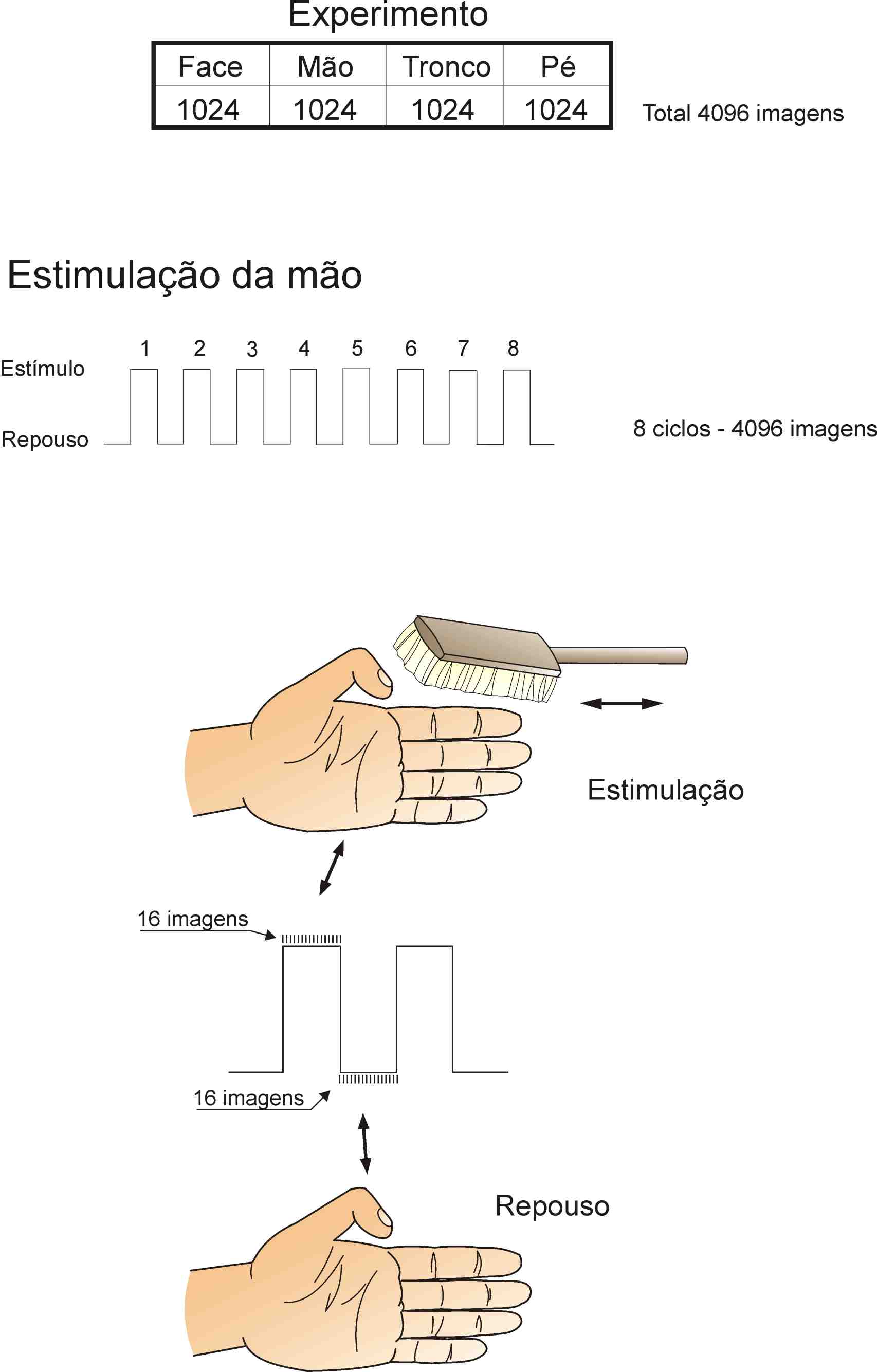
 In this experiment, we will try to describe the areas of brain cortex which are responsible for the treatment of tactile information (touch). Therefore, parts of the experiment will be programmed to stimulate segments of skin in the hand, trunk and feet (Fig. 1 - top). The visualization of the areas of cortex activated by sensorial stimulation will allow the localization and topographical organization of some sthetic (touch) areas of human beings.
In this experiment sets of images (images of eight brain slices at different heights) will be acquired during the period of stimulated alternated with sets of images at rest. Figure 1 shows an example of three slices. Thus, in each cycle of stimulation and rest we would acquire eight sets of sixteen images of eight slices of the brain in a total of 128 images. During one phase of the experiment the stimulation of one region of the body, for instance, fingers, would berepeated eight times, resulting in a set of 1,024 images to each body area studied (Fig. 2). The total experiment, studies four different regions of the body including regions in the hand, face, trunk and foot. The data consist of 4,096 images taken under two basic situations: stimulation and rest.
 Having acquired the images under two sets of situation it matters to identify which areas of the brain showed the same variation in magnetic signal that correlates with the function rest/stimulation (Time course screen - Fig 3). The first method developed with this aim was an averaging technique during stimulation, followed by the subtraction of the average during rest. In this method, the image is divided into pixels ("little squares in the screen") where each element is attributed a value of the magnetic resonance intensity. The subtraction operation made pixel by pixel shows small areas of differential rest/stimulation signals. The difference in resonance can then be standardized and coded in colors to be superimposed to the structural image of the slices under investigation (slice on the right in Fig. 3) Another method we use is the correlation between the magnetic signal of each pixel and the stimulation line. This correlation method allows a more general analysis of the stimulation/rest correlation, besides allowing multiple correlation analysis when the experiment is conducted with more than two variables. The methodology used involves the application of statistical tools that correlates the variation of intensity of the signal in each voxex (3-D pixel) of the image with a calibration curve determined according to the periods of stimulation and control (cross-correlation analysis). The threshold of correlation is set to 0.7, that is, all the pixels with correlation less than 0.7 will be discarded. Therefore we will get a map of all pixels that are above the threshold limit. To increase the specificity of the study, these maps will be treated with another tool called the grouping technique (Fig. 4). Consequently, only the areas where three or more neighboringpixels exceed the coefficient are considered as real activation.This kind of treatment discards the vast majority of pixels with random correlation, thus directing the analysis and consolidating the results.
Tactile Sensorial Areas in Human Cortex
 The results shown in Figure 1 demonstrate several somesthesic areas in humans.Several focus of activation in the s1 Area (Broadman's 3-1-2 areas)indicate the existence of further two somesthesic areas in the pre-central depression. Besides these, two other areas were discovered: the so-called S2, located in Brodaman's 43 area) and another called insular somesthesic area, situated in the insular cortex at the junction of pre-central twist and parietal cortex.
Binder JR, Rao SM, Hammeke TA, et al. Lateralized human brain language systems demonstrated by task subtraction functional magnetic resonance imaging. Arch Neurol, vol.52, 1995; 593-601.
Ogawa S, Menon RS, Tank DW, et al. Functional brain mapping by blood oxigenation level-dependent contrast magnetic resonance imaging. Biophys J 1993; 64:803-812.
Ogawa S, Lee TM, Kay AR, et al. Brain magnetic resonance imaging with contrast dependent on blood oxigenation. Proc Natl Acad Sci USA 1990; 94:68-78.
Ricardo Gattass, MD, PhD
E-mail: rgattass@chagas.biof.ufrj.br,rgattass@abc.org.br
Researcher and Full Professor, Institute of Biophysics,
Federal University of Rio de Janeiro.
Member, National Academy of Sciences, Section of Biological Sciences
Full member since 20/12/93