 Neurons and Synapses
![]() The Microcosm Arrives
The Microcosm Arrives
![]() The Discovery of Neurons and Its Processes
The Discovery of Neurons and Its Processes
![]() The Neuronal Doctrine
The Neuronal Doctrine
![]() The Discovery of the Synapse
The Discovery of the Synapse
![]() Chemical Transmission
Chemical Transmission
![]() To Know More
To Know More
![]() The Author
The Author
The second quarter of the 19th century was a period where young neuroscience was ripe for a sucession of seminal discoveries about the brain. In Great Britain, Italy, Germany and France, new and exciting lines of research were being pursued. By the start of the next century, they would begin to converge into a powerful scientific synthesis of our knowledge about this most complex organ.
The search for localization of nervous and mental function received a mighty push by Pierre Flourens, a distinguished French physiologist, who, in his quest to disallow Franz Joseph Gall´s equivocal ideas about localization, was able to prove, from 1825 to 1827, that the main divisions of the brain were, indeed, responsible for quite different functions.
In another field of inquiry, Carlo Mateucci, Emil du Bois-Reymond, Julius Bernstein and their followers were using increasingly sophisticated physical apparatuses, toward the end of the 1850s, to investigate the electrical nature of nerve impulses and the new model of functioning of the nervous system. This model had, by the able hands of Luigi Galvani, substituted the old vitalistic, "animal spirits", hydraulic model, respected since the ancient Greek philosophers, and of which the philosopher René Descartes was the most recent and notable proponent.
However, nothing was known yet about the fine organization of the nervous system. The macroscopic structures of the spinal chord, the nerves and the brain where already fairly well known, but its inner workings were a complete mistery. The distinction between white and gray matter was almost everything that science knew about the nervous tissue itself. In fact, until 1870, anatomists, even armed with powerful microscopes, did not know the cellular structure of the nervous system, and referred to what they saw as "globules" instead of cells. The neuron received its name in 1891, only!
But, around the same time, a new line in the study in the anatomy of the nervous system was starting to bring light to these baffling aspects. It was brought about by the able application of a new and powerful instrument of discovery, the compound optical microscope, together with an array of associated techniques for fixing, cutting and staining the elusive, fragile, almost featureless neural tissue. Access to the most mysterious of all biological spheres, the microcosm, was now granted to curious scientists.
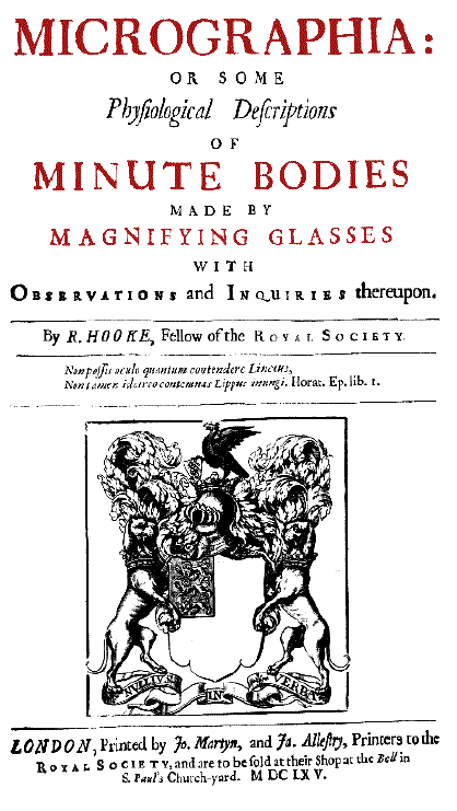
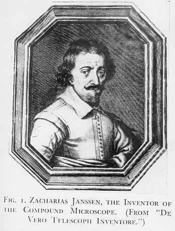
 The microscope was first built in 1595 by Hans and Zacharias Jansen (1588-1631) in Holland (see figure).
Later, it was perfected in the 17th century in several countries, including by Robert Hooke (1635-1703), in England
but most notably by a Dutchman, Anton van Leeuwenhoek (1632-1723). Hooke, after examining thin pieces of cork,
discovered it had a honeycombed structure, and used for the first time the word "cell" to describe its
smaller elements. Using a much improved microscope, with a monocular eyepiece, a wooden tube, a stage for holding
a specimen, and a glass globe full of water to concentrate light onto it, Hooke produced marvelous illustrations,
which were published in 1667, in his famous book Micrographia,
which fired the imagination of his contemporaries, including van Leeuwenhoek.
The microscope was first built in 1595 by Hans and Zacharias Jansen (1588-1631) in Holland (see figure).
Later, it was perfected in the 17th century in several countries, including by Robert Hooke (1635-1703), in England
but most notably by a Dutchman, Anton van Leeuwenhoek (1632-1723). Hooke, after examining thin pieces of cork,
discovered it had a honeycombed structure, and used for the first time the word "cell" to describe its
smaller elements. Using a much improved microscope, with a monocular eyepiece, a wooden tube, a stage for holding
a specimen, and a glass globe full of water to concentrate light onto it, Hooke produced marvelous illustrations,
which were published in 1667, in his famous book Micrographia,
which fired the imagination of his contemporaries, including van Leeuwenhoek.
Van Leeuwenhoek used his new instrument, which was tenfold more potent than Hooke´s (he reached the amazing power of 300 times with a single lens!) to discover startling microscopic things, such as protozoa and spermatozoa, which thus far were completely unknown to science, or to discover the microscopic structure of known things, such as fleas and plant leaves. Van Leeuwenhoek had even been able to slice specimens of a cow's optical nerves in 1674, and observe its longitudinal fibrous internal structure. He was perplexed to see that they were not hollow tubes, as the prevailing theory of the time, such as that defended by René Descartes, proposed.
|
|
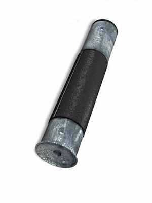
primitive one-lens microscope |
|
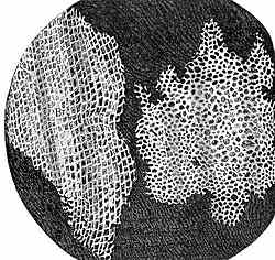
|
showing a longitudinal and a cross section of a nerve. |
by Robert Hooke |
 |
 Unfortunately,
these early microscopes were rudimentary, low-power devices, and suffered, along the whole of the 18th century,
of severe restrictions to allow the investigation of natural objects which were much harder to visualize, such
as bacteria and the cells of organic tissues. In fact, the microscope was considered a dilettante's or amateurish
instrument, and scientists shied away from its serious use, lest they ran the risk of being ridiculed by their
peers!
Unfortunately,
these early microscopes were rudimentary, low-power devices, and suffered, along the whole of the 18th century,
of severe restrictions to allow the investigation of natural objects which were much harder to visualize, such
as bacteria and the cells of organic tissues. In fact, the microscope was considered a dilettante's or amateurish
instrument, and scientists shied away from its serious use, lest they ran the risk of being ridiculed by their
peers!
In the beginning of the 19th century (1824), however, the invention of the achromatic lens and the development of the compound optics microscope, allowed anatomists to visualize with increased sharpness very small structures, and cells in many parts of the body were documented, because these new improved microscopes permitted a higher power of magnification without distortions (the so-called spherical aberration) and separation of colors in the image (chromatic aberration). Powerful light sources and precision mechanical stages and focusing screws added to the microscope usefulness as a scientific instrument.
Furthermore, better techniques for hardening ("fixing") with alcohol,
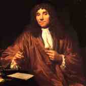
a discovery made in 1805 by Johann Christian Reil (1759-1813) and then sectioning the fragile brain sections came into existence.
Another important development in the histological technique was staining, or
adding color and opacity to the usually translucent and colorless brain cells. Although van Leeuwenhoek experimented
a bit with wine spirit (alcohol) and a saffron stain to wet his specimens (particularly muscles) before observing
them at the microscope, these techniques were perfected only much later, in the second half of the next century.
As we will see below, they became a most important factor in the scientific development of microscopy of neural
tissue, and eventually led to two Nobel awards in 1906, the very first in neuroscience.
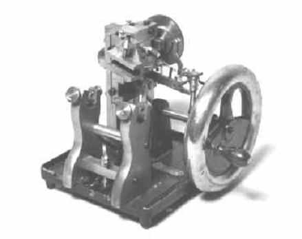
Rotary microtome (1905)At first, histologists sectioned specimens by hand, using sharp razors.
Later, around 1790, the first microtome was devised. It consisted of a wooden device with a berth to accomodate
the specimen and a flat surface to slide the razor against the specimen surface, thus increasing blade purchase,
regularity and thinness of the slice. The term microtome was given to these instruments by Charles Chevalier, who perfected it around
1825. Finally, around 1870, precision mechanical devices were developed. They consisted of a metal stage holding
a parafin or celloidin block with the embedded piece of the tissue to be sectioned and a mechanical swing holding
the blade in precise alignment with the stage. The blade is swung against the specimen´s surface by either
a rotary or a rocking mechanism. Thus, serial sectioning was achieved, a very important technique for tracing neurons
across the three-dimensional space of the brain.
Next...
Prof. Renato
M.E. Sabbatini, PhD is a neuroscientist and a specialist in medical informatics,
holding a doctoral degree in neurophysiology by the University of São Paulo, Brazil, and a post-doctoral
fellowship at the Max Planck Institute for Psychiatry, in Munich, Germany. He is the director of the Center for
Biomedical Informatics and associate professor and chairman of medical informatics at the Faculty of Medical Sciences,
both at the State University of Campinas, Brazil. Email: sabbatin@nib.unicamp.br


