
 Why
Einstein Was a Genius?
Why
Einstein Was a Genius?
By: Silvia Helena Cardoso, PhD
We always suspected that something physically extraordinary must have made Albert Einstein smarter than the rest of us. His contributions changed our conceptions of space, time, and the very nature of reality, and his ideas have left their mark on nearly every aspect of modern physics, from the subatomic to the cosmological.
Einstein even said himself that one of the keys to his intelligence was in his ability to visualize the problems he was working on and then translate those visual images into the abstract language of mathematics. In fact, one of the most famous examples is his special theory of relativity, which, as the story goes, he developed out of day dreams of what it would be like to ride through the universe ona beam of light.
When Einstein died in 1955 at the age of 76, his body was cremated. Before that, Dr. Thomas Harvey, a pathologist who performed the autopsy, took the brain home with him. Some parts of the brain were given to scientists to be used for scientific studies. The brain was not heard from again until 1978 when the reporter Stephen Levy tracked it down to Harvey's office in Kansas. According to Levy, Einstein's brain was being stored in Harvey's office inside two jars. Most of the brain, except for the cerebellum and parts of the cerebral cortex, had been sectioned (sliced). Dr. Harvey's preliminary examinations had found nothing unusual about the anatomical structure of Einstein's brain.
One of the scientists who got a part of Einstein's brain was Marian Diamond, a prominent Berkeley professor.
 She
and her team counted the number of neurons and glial cells in Einsteins
brain: area 9 and area 39 of the cerebral cortex on the right and left
hemisphere. Area 9 is located in the frontal lobe (prefrontal cortex) and
is thought to be important for planning behavior, attention and memory.
Area 39 is located in the parietal lobe and is part of the "association
cortex." Area 39 is thought to be involved with language and several other
complex functions. The ratios of neurons to glial cells in Einsteins brain
were compared to those from the brains of 11 men who died at the average
age of 64.These scientists reported that Einstein's brain appeared to have
a higher percentage of glial cells, the cells that support and nourish
the network of neurons (1). The group concluded that the greater number
of glial cells 'oligodendroglia' -- helper cells that speed neural
communication -- per neuron might indicate the neurons in Einsteins brain
had an increased "metabolic need" - they needed and used more energy. In
this way, perhaps Einstein had better thinking abilities and conceptual
skills. However, it is important to note that the areas 9 and 39 make important
connections with many other areas of the brain and complex behavior is
the result of many areas acting together.
She
and her team counted the number of neurons and glial cells in Einsteins
brain: area 9 and area 39 of the cerebral cortex on the right and left
hemisphere. Area 9 is located in the frontal lobe (prefrontal cortex) and
is thought to be important for planning behavior, attention and memory.
Area 39 is located in the parietal lobe and is part of the "association
cortex." Area 39 is thought to be involved with language and several other
complex functions. The ratios of neurons to glial cells in Einsteins brain
were compared to those from the brains of 11 men who died at the average
age of 64.These scientists reported that Einstein's brain appeared to have
a higher percentage of glial cells, the cells that support and nourish
the network of neurons (1). The group concluded that the greater number
of glial cells 'oligodendroglia' -- helper cells that speed neural
communication -- per neuron might indicate the neurons in Einsteins brain
had an increased "metabolic need" - they needed and used more energy. In
this way, perhaps Einstein had better thinking abilities and conceptual
skills. However, it is important to note that the areas 9 and 39 make important
connections with many other areas of the brain and complex behavior is
the result of many areas acting together.
The most recent finding on Einstein's
brain was published on June, 1999. Scientists have found that one part
of his brain was indeed physically extraordinary. A team of department
of Psychiatry and Behavioural Neurosciences, from the Faculty of Health
Sciences at McMaster University compared anatomical measurements of Einstein's
brain with those of brains of 35 men and 50 women who had normal intelligence.
In general, Einstein's brain was similar to the other brains except for
one area called the inferior parietal region. Because of extensive development
of this region on both sides of his brain, his brain was 15% wider than
other brains studied. "Visuospatial cognition, mathematical thought, and
imagery of movement are strongly dependent on this region," the researchers
note. This unusual brain anatomy may explain why Einstein tackled scientific
problems the way he did.
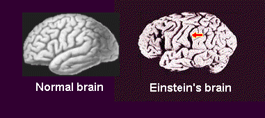
Normal brain - contains a sulcus called the parietal operculum and the inferior parietal lobe; the latteris the seat of mathematical and visual reasoning Einstein brain - was no longer than most, but the parietal operculum region was missing. This allowed the inferior parietal lobe to grow 15% wider than normal |
In addition, Einstein's brain was unique
in that it did not have a groove, called a sulcus, that normally runs through
part of this area. The researchers speculate that the absence of the groove
may have allowed more neurons in this area to establish connections between
each other and work together more easily, possibly creating an "extraordinarily
large expanse of highly integrated cortex within a functional network."The
findings, the researchers conclude, suggest that differences in people's
ability to perform certain cognitive functions may be due in some degree
to physical differences in the structure of the regions of their brains
that mediate those functions.
Witelson theorized that the partial absence of the groove in Einstein's brain may be the key, because it might have allowed more neurons in this area to establish connections between one another and work together more easily. |
Not only was Einstein's inferior parietal region unusually bulky, but a feature called the Sylvian fissure was much smaller than average. Without this groove that normally slices through the tissue, the brain cells were packed close together, permitting more interconnections - which in principle can permit more cros-referencing of information and ideas, leading to great leaps of insight.
In conclusion, Although these results are interesting, it remains to be seen if every brilliant physicist and mathematician will have this same anatomy. Looking at the brains of living geniuses may be easier than it was with Einstein. In the past, a person's brain anatomy could be studied only after his or her death, but modern technology, such magnetic resonance imaging (MRI) and positron emissiontomography (PET), allows scientists to observe the brain at work within the living body. With this technology, it may prove quite possible to observe, not only differences in brain structure, but also the actual amount ofactivity taking place in those structures. For instance, if Einstein'sbrain had been studied with this technology, scientists could have observed the larger, unique parietal lobes and looked for activity in those areas as the physicist thought about his theories. Moreover, the study did not investigate how neurons in these brains were connected and of course, could not tell if there were differences in the way the neurons functioned.
References
The Author

Silvia Helena
Cardoso, PhD. Psychobiologist, master and doctor in Sciences,
Founder and editor-in-chief,Brain
and Mind Magazine, State University of Campinas.