![[Brain & Mind Header and Navigation Bar]](../../arttec_i.jpg)
![[Siemens Exact PET Scanner]](exact.gif)
Physicians and researchers have a new and wonderful tool to understand how the brain works in health and disease. It's the PET scanner, a kind of computerized tomography machine which is able to pinpoint in brilliant color the regions in the brain where nerve cells are working during a particular mental task. PET means Positron Emission Tomography, and is based on a host of exciting new technological developments in nuclear medicine and digital imaging of the brain, which is already revolutionizing the way we can study the function of the nervous system in animals and patients alike. In this article, you will know more about PET and its some of its recent uses in the neurosciences.
![[Marcus Raichle's PET image of training brain]](petrain2.gif) A
good example of the fantastic imaging capabilities of PET is shown in the
images at the left, made by Dr. Marcus
Raichle, at the Neuroimaging Lab
or the Washington University School of Medicine, St Louis, USA.
A
good example of the fantastic imaging capabilities of PET is shown in the
images at the left, made by Dr. Marcus
Raichle, at the Neuroimaging Lab
or the Washington University School of Medicine, St Louis, USA.
They were taken under two different conditions. In the first one (uppermost image), an individual was hearing a text, in order to learn a new language task. The color map shows the regions of the brain which were activated by this task, in other words, where there were cells working more than in their resting state, with a higher metabolism (using more energy and more blood flow). The PET machine shows the degree of activity in several tones of color, like in a rainbow. Yellow and red regions are "hotter", that is, they indicate a higher cell activity. Blue and black regions show decreased activity or none at all. While obtaining this image, the patient was still unpracticed at the language learning task. The highest brain activities are shown in an area called temporal lobe, responsible for the hearing perception, and in another area called prefrontal cortex, responsible for understanding language.
In the second condition (lowermost image), the same individual has now learned the language task and is spelling out. You can easily see in the color map that two different regions of the brain were activated in each condition. Now the activity is concentrated in the area of the cortex which is responsible for the motor control of voice, the so-called area of Broca, so named because it was discovered by a French physician named Paul Broca, in the turn of the century. Thus, the functional map obtained with PET closely corresponds with what we know about the brain's functional neuroanatomy, discovered by other methods. The difference here is that we can actually obtain a real-time image of brain function.
The PET scan can be used for a whole range of experimental and clinical studies of the brain. A veritable explosion of new research on the functions of the brain has been caused by a wider availability of PET equipments around the world (they are very expensive, several million dollars each, and are also costly to operate). Today (beginning of 1997, there are more than 150 installations around the world.
How we will see later, PET can be used to study in-vivo (i.e., in live subjects) blood flow, blood volume, oxygen consumption, tissue pH (acidity), glucose utilization and the activity of drug receptors in brain cells. IT shows the localization of function, rather than anatomy, so that it will give positive results were other methods, such as magnetic resonance tomography (MRI), fail. When the alteration in the structure which can be seen in a CT scan (using x-rays) also produces a metabolic alteration, such as a brain tumor, the PET image will show that, too.
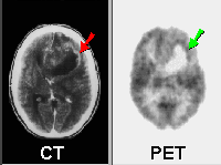
![[PET in Epilepsy]](epilepsy.gif) The
availability of PET has shed a new, exciting light onto many brain diseases
and pathological conditions. For example, the image at the right shows
the abnormally high activation of the right part of the brain of a patient
during an epileptic seizure (the yellow and magenta patches). What you
see here is a series of transversal sections, i.e, the apparatus gets images
like horizontal slices through the brain. Each slice is made at a different
level, a few centimeters from the preceding one. This allows the physician
to locate with remarkable precision where the changes induced by the disease
are taking place. This information can be used to understand better what
is going on, to make a better diagnosis, or even to allow a surgical intervention,
if necessary.
The
availability of PET has shed a new, exciting light onto many brain diseases
and pathological conditions. For example, the image at the right shows
the abnormally high activation of the right part of the brain of a patient
during an epileptic seizure (the yellow and magenta patches). What you
see here is a series of transversal sections, i.e, the apparatus gets images
like horizontal slices through the brain. Each slice is made at a different
level, a few centimeters from the preceding one. This allows the physician
to locate with remarkable precision where the changes induced by the disease
are taking place. This information can be used to understand better what
is going on, to make a better diagnosis, or even to allow a surgical intervention,
if necessary.
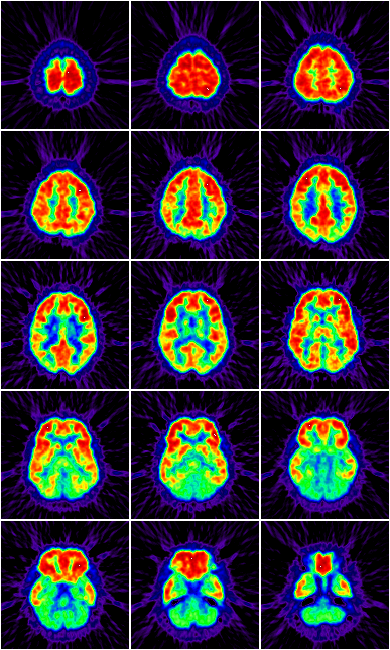
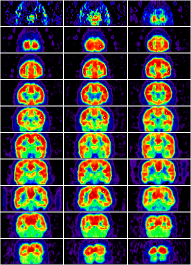
Usually a single study is comprised by a series of transverse or frontal sections of the
brain.
![[PET of Parkinson's transplantation]](pettrans.gif) Another
interesting example of PET's usefulness is shown in this study of a patient
with Parkinson's disease, made in the PET installation of the Crump
Institute for Biological Imaging, University of California at Los Angeles.
This disease is characterized by many neurological alterations, such as
muscle rigidity, tremor of the hands, speech slurring, difficult gait,
etc. It is caused by a widespread lesion of some areas of the brain, called
basal ganglia, which then produce less endogenous chemical substances,
such as DOPA, which are necessary for the motor functions.
Another
interesting example of PET's usefulness is shown in this study of a patient
with Parkinson's disease, made in the PET installation of the Crump
Institute for Biological Imaging, University of California at Los Angeles.
This disease is characterized by many neurological alterations, such as
muscle rigidity, tremor of the hands, speech slurring, difficult gait,
etc. It is caused by a widespread lesion of some areas of the brain, called
basal ganglia, which then produce less endogenous chemical substances,
such as DOPA, which are necessary for the motor functions.
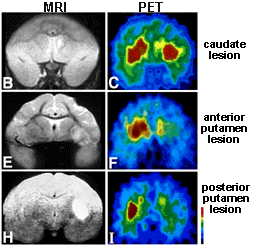
In the sequence of image, a PET machine was used to make an image of the brain showing where there is a higher concentration of DOPA (the bean shaped regions in red and green are a part of the basal ganglia). The uppermost image shows a normal brain, the middle one the brain of a patient with Parkinson (you can see how the brain concentration of DOPA has decreased(, and the third shows the brain of the same patient after receiving an implant of DOPA-secreting tissue (tha concentration is nearly normal again). This is fabulous stuff, making possible a new window into the brain.
![[PET of a Wada Test]](wadasm.gif) PET
is also a very useful tool for investigating cognitive functions (for example,
language, conscience, learning, sensory processing, etc.). For example,
the Wada Test is used to check language and memory functions. The
subject receives an injection of a short-acting barbiturate, amobarbital,
injected into one of the carotid arteries. The hemisphere of the same side of
the injection is inactivated, and the language and memory tests are performed.
As the image in the right shows, only one hemisphere is activated.
PET
is also a very useful tool for investigating cognitive functions (for example,
language, conscience, learning, sensory processing, etc.). For example,
the Wada Test is used to check language and memory functions. The
subject receives an injection of a short-acting barbiturate, amobarbital,
injected into one of the carotid arteries. The hemisphere of the same side of
the injection is inactivated, and the language and memory tests are performed.
As the image in the right shows, only one hemisphere is activated.
Finally, two last examples of PET capabilities:
![[PET of developing brain [UCLA]]](petdev.gif) |
![[PET of a brain tumor]](pettum.gif) |
| These images, also done at the Crump Institute at UCLA, show the increase of brain activity which accompanies the growth of the brain, in the same patient, from the age of 1 to 12 months. This can be used, for instance, to pinpoint developmental problems in children, much earlier than other tests would show. | This image shows a brain tumor. The PET is very useful not only to detect a tumor when it's in the initial stages of growth, making the treatment more effective in eradicating it, but also to detect it's type, malignancy and spread, without the need to open the patient's brain to carry out a risky biopsy |
PET is now being used by physicians to provide valuable information in many neurological diseases such as Alzheimer's disease, dementias, Parkinson's disease, Huntington's disease, and Down's Syndrome. It is also being extensively used in Psychiatry, because PET is very sensitive to biological brain alterations during episodes of schizophrenia, depression and other disorders.
In conclusion it has a brilliant future in medicine, psychology, psychiatry, and in the neurosciences in general, for studying the relation between structure and function.
![[Books]](books2.gif)
Also, do not miss the excellent article "Visualizing the Mind", by Dr. Marcus Raichle, in the April 1994 issue of Scientific American magazine. Other books you can order on-line are Images of Mind (W.H. Freeman Publ, 1994) and Mapping the Brain and Its Functions (National Academy Press, 1991). Two interesting scientific journals are Human Brain Mapping and NeuroImage.
| Renato M.E. Sabbatini is a neuroscientist and specialist in Biomedical Informatics, with a doctoral degree by the University of São Paulo and a postdoctoral fellowship in the Max Planck Institute for Psychiatry, Munich, Germany. He is the current director of the Center of Biomedical Informatics and professor of Medical Informatics of the Faculty of Medicine of the State University of Campinas, Brazil. Email: sabbatin@nib.unicamp.br |
Center for Biomedical Informatics
State University of Campinas, Brazil
Copyright © 1997 State University of Campinas