![[Brain & Mind Header and Navigation Bar]](../../arttec_i.jpg)
The PET scanner is a new kind of medical instrument which is radically different from the tools which the physicians had to make images of the brain in a non-invasive way, that is, without having to open the skull in order to peer inside or to actually take samples of brain tissue.
And why PET is different ?
The first non-invasive technique to get images from the brain was a big revolution in itself. It was the radiography, invented by a German physicist named Wilhelm Röntgen, in 1896, and it used x-rays, an invisible eletromagnetic radiation discovered also by him. To Röntgens astonishement, x-rays were able to penetrate the body, as it were transparent, and to produce a negative photograph of the body's interior, showing with startling detail the bones, cavities and other anatomical structures on its path. Röntgen could see immediately the medical value of his discovery, and the first radiograph ever made was from his wife's hand.
However, x-rays can shown only the anatomical structures, and nothing else. The function of these structures could be inferred from anatomical changes, but only when they happened. Enlargement, movement and flow of substances could be observed in some selected organs (for example, the heart or the intestines, or, by using some liquids which are opaque to the x-rays, named contrasts), but not much more. Since the brain does not move, radiographys is of little value to study function, particularly normal function.
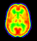
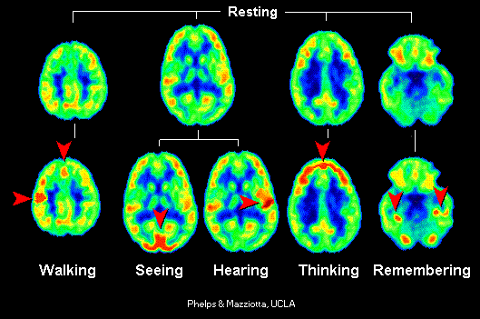
The immense value of PET for studying normal brain function was highlighted by several path-breaking investigations. The UCLA researcher Michael E. Phelps, for instance, was one of the first to show in striking detail how different parts of the brain are activated when performing mental tasks such as hearing, reading, talking, thinking, etc.
 The
first PET scanners had a small number of radiation sensors to build
the image, and they could do only a slice at a time. The slices were also
very thick. Thus, the images obtained with the PET had a low quality and
definition. It was impossible to get the finer details of localization
of function in the brain, so their clinical usefulness was quite limited,
as compared with modern models.
The
first PET scanners had a small number of radiation sensors to build
the image, and they could do only a slice at a time. The slices were also
very thick. Thus, the images obtained with the PET had a low quality and
definition. It was impossible to get the finer details of localization
of function in the brain, so their clinical usefulness was quite limited,
as compared with modern models.
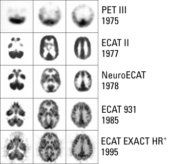 Thisfigureshows
the evolution of image quality from the first PET scanner, available in
1975, to the latest and most sophisticated model, ECAT Exact HR+. You can
easily see how the finer detais of internal brain structures can be visualized
much better in the latest model. This improvement has been achieved with
a larger number and better radiation sensors, better computer programs
and the possibility of getting several slices at the same time (using many
rings of sensors). All models of PET machines depicted here were produced
by the Siemens biomedical equipment manufacturer, which has installed the
majority of PET scanners in the world.
Thisfigureshows
the evolution of image quality from the first PET scanner, available in
1975, to the latest and most sophisticated model, ECAT Exact HR+. You can
easily see how the finer detais of internal brain structures can be visualized
much better in the latest model. This improvement has been achieved with
a larger number and better radiation sensors, better computer programs
and the possibility of getting several slices at the same time (using many
rings of sensors). All models of PET machines depicted here were produced
by the Siemens biomedical equipment manufacturer, which has installed the
majority of PET scanners in the world.
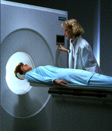
 Modern
PET scanners are very expensive and sophisticated pieces of equipment.
They are also much easier to install and to operate, and have many new
capabilities which clinicians use with advantage to perform many feats
of brain imaging, such as a higher speed in obtaining results. For example,
as shown here, they can be used to produce movies of parts of the body
Modern
PET scanners are very expensive and sophisticated pieces of equipment.
They are also much easier to install and to operate, and have many new
capabilities which clinicians use with advantage to perform many feats
of brain imaging, such as a higher speed in obtaining results. For example,
as shown here, they can be used to produce movies of parts of the body
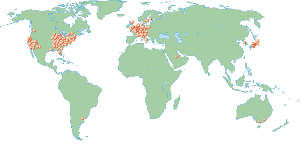 Since
PETs are so expensive (actually, it's rather the whole installation, including
the cyclotron to produce the radiopharmaceuticals which adds to the cost),
there are only about 150 installations around the world, which are highly
concentrated on the USA (particularly in the East and West coasts), Europe
(particularly in the Anglo-Saxon countries and France) and in Japan. In
the Southern hemisphere, only Australia and Argentina have a very small
number of PET facilities.
Since
PETs are so expensive (actually, it's rather the whole installation, including
the cyclotron to produce the radiopharmaceuticals which adds to the cost),
there are only about 150 installations around the world, which are highly
concentrated on the USA (particularly in the East and West coasts), Europe
(particularly in the Anglo-Saxon countries and France) and in Japan. In
the Southern hemisphere, only Australia and Argentina have a very small
number of PET facilities.
Image credits: The Crump Institute for Biological Imaging, Department of Pharmacology, University of California at Los Angeles. CTI and The History of Medicine CD-ROM,
From: The PET Scan: A New Window Into the
Brain
By: Renato M.E. Sabbatini,
PhD
In: Brain & Mind Magazine, March 1997.